32. ICOSIM Weekly

1. Free ICOSIM Webinar on 21.02.24

2. New Publication: Is There a Question Deficit in Implantology?

3. “Do Not Miss” Dates 2024

Dear friends of ICOSIM and
jawbone osteoimmunology,


The new year 2024 starts with full power!

1. Free ICOSIM Webinar on 21.02.24


2. Next Article: Is There a Question Deficit in Implantology?


Who in implantology considers the “immunological sustainability” of invasive procedures? In this context, an implant significantly interacts with osteoimmunological processes related to jawbone quality, especially when it goes beyond simple drilling and post placement. Does it matter for the local quality of osseointegration whether the material is titanium or ceramic? Or does individual material hypersensitivity play a role in this?

These systemic-immunological questions about osseointegration and osteoimmunology, beyond peri-implantitis and mechanical BIC, are addressed for the first time in our latest PubMed-indexed study using radiation-free trans-alveolar ultrasound sonography (US) and precise bone density measurement (CTU) with US/CTU (comparable to Hounsfield units):

Lechner J, von Baehr V, Notter F, Schick F.


Osseointegration and Osteoimmunology in Implantology: Assessment of the Immune Sustainability of Dental Implants Using Advanced Sonographic Diagnostics: Research and Case Reports.

Journal of International Medical Research. 2024;52(1). doi:10.1177/03000605231224161

For a free download (beware, it’s a 28-page PDF!) and an introduction to scientific work with ultrasound sonography, visit www.cavitau.de.

Our ultrasound sonography provides the following overview:

  • US/CTU measurements indicate reduced Bone-to-Implant Contact (BIC) and diminished osseointegration in Ti-Stim-positive collectives compared to Ti-Stim-negative collectives. Additionally, Ti-Stim-positive groups exhibit higher inflammation levels.
  • CTU measurements reveal better osseointegration and an eightfold higher BIC for ceramic implants compared to titanium implants.
  • Multiplex analyses of RANTES/CCL5 expression demonstrate approximately 20-fold increased cytokine expression around titanium implants with bone marrow defects.

For more information on osteoimmunology in dentistry and to join the International College of Maxillo-mandibular Osteoimmunology (ICOSIM e.V.), visit www.icosim.de.

3. “Do Not Miss” Dates 2024

a) Tissue Master Congress: CaviTAU and lecture: March 14-15 in Nuremberg


b) Not only the Bundeswehr, but also CaviTAU® in Lithuania! 


c) Craving for Sunshine: Barcelona, April 2024

I have the occasion to lecture on the “XXI National Congress of Kinesiology”, which will be held at the Espai Pujades 350 in Barcelona on April 11, 12 and 13, 2024.

My presentations are scheduled for

Thursday, April 11 at 3:00 p.m. – 7:00 p.m. with the title “Pre-Congress Course: Integrative Oral Medicine and Osteoimmunology” and

Saturday , 12 april at 13.15 p.m – 13:45 pm with the title “Unattended systemic diseases of mandibular cause diagnosed by ultrasound”

Congress website: https://www.sekmo.es. Instagram link of my intervention planned https://www.instagram.com/p/C2ejmeyIdUu/


I remain with collegial greetings until the next ICOSIM Weekly.

Your Hans Lechner.

31. ICOSIM Weekly

1. New publication: Does X-ray lead dentists to the wrong pathology?

2. Call for multicenter study

Dear friends of ICOSIM and
jawbone osteoimmunology,


First of all, I wish you a Happy New Year 2024!

And I am also happy to announce some good news:

1. New article on CaviTAU® and RANTES/CCL5 with the central question


Does X-ray lead dentists to the wrong pathology? 

In collaboration with the scientific institutes of the University of Lisbon/Dr. Joana Vasconcelos e Cruz, our Munich Research Team (Lecher/Notter/Schick) has presented another scientific publication in the PubMed-indexed Journal of Inflammation Research:

Comparison of cytokine RANTES/CCL5 inflammation in apical periodontitis and in jawbone cavitations – Retrospective clinical study


The central statements of your current publication are:

a) Even the apical granulomas/apical periodontitis visible in the X-ray express RANTES/CCL5.

b) However, the FDOK areas that are not detectable in the X-ray exhibit a 3x higher inflammation pattern of RANTES/CCL5.

c) The actual pathogenic chronic inflammatory triggers of the immune diseases caused by RANTES/CCL5 overexpression (such as rheumatism, cancer, neuroinflammation, etc.) are regularly not treated by dentists, as they are only detectable with ultrasound sonography using CaviTAU®.

d) Conclusion: The fixation on the X-ray image prevents the treatment of systemic immune diseases in dentistry; a complementary application of ultrasound sonography is necessary from an integrative medical perspective for patient protection.


The graphic shows the unequal distribution of RANTES/CCL5 expressions in pg/mL:


Here is the translation of your message to English: “An illustration of the 4 case reports shows the difference in radiological presentation of a chronically inflammatory bone marrow defect compared to the imaging of ultrasound sonography CaviTAU® and the RANTES/CCL5 overexpression of 3,928 pg/ml compared to the norm of 149.9 pg/ml.

Such articles in medical journals are of invaluable value to our practice success in distinguishing us from less advanced colleague practices!


2. Call for a multicenter study


For another multicenter study on the CCL5/CCR5 axis and breast cancer using the reference standard RANTES/CCL5 discovered by us, we are looking for colleagues who

a) perform FDOK with a RANTES/CCL5 multiplex analysis at IMD (following the example of Hans Lechner) for forensic control

and

b) perform these FDOK operations on patients with breast cancer


Is this applicable? Then please contact ICOSIM project leader Hans Lechner immediately!


I am looking forward to hearing opinions and questions from our ICOSIM colleagues and remain with collegial greetings until the next ICOSIM Weekly.

Your Hans Lechner.

30. ICOSIM Weekly

1. Newspaper article: successful treatment of trigeminal neuralgia, facial pain and Parkinson’s disease

2. Case of the week: Trigeminal neuralgia again despite Janetta surgery!

Dear friends of ICOSIM and
jawbone osteoimmunology,


1. Article about our successes with patients

 
I am pleased to be able to share a nice surprise with you: A great article has appeared in the Swedish newspaper TF-bladet about our successes with patients that we have been able to achieve with the help of CaviTAU®. The reference to the success of “Ultrasonography Guided Jawbone Detox” in the treatment of trigeminal neuralgia and facial pain and to a case of Parkinson’s disease that we cured with CaviTAU® and Jawbone Detox is journalistically perfectly presented!

Such articles in medical lay portals are of course invaluable for the success of our practice!

Here you can download the article as original in Swedish or as translated English version:


2. The case of the week: Trigeminal neuralgia despite “Janetta surgery” at the base of the skull


An approximately 55-year-old female patient presents to our practice clinic with trigeminal neuralgia in the upper right jaw. Her history is so interesting because after 15 years of constant neuralgia she found freedom from pain after a Janetta operation on the right. However, this only lasted for around 18 months, as she has now had the same trigeminal neuralgia again for 6 months. Our CaviTAU® image clearly shows why the pain in the upper right has returned: The osteoimmunologic inflammation in the alveolar bone OK right is still active!


Questions about the case:

a) Why do neurologists rush straight to Janetta surgery at the base of the skull instead of starting diagnosis and therapy at the peripheral nerves in the jaw area?

b) Should we then be surprised that the initial success of the Janetta operation disappears if the chronic inflammatory FDOJ irritation from the periphery persists?

c) Of course, a dentist has already offered to “pull the teeth” in the past: without correcting the osteoimmunologically dysregulated jawbones, this would only have led to a widespread negative assessment by the neurologist: “Just don’t pull teeth!”


I look forward to hearing opinions and questions from our circle of ICOSIM colleagues on this topic and remain with best wishes for the festive season and best regards from my colleagues until the next ICOSIM Weekly in the New Year 2024!

Your Hans Lechner.

29. ICOSIM Weekly

1. Excerpt from our scientific study in the Journal of International Medical Research

2. FDOK documentation with 69 images and histologies

Dear friends of ICOSIM and
jawbone osteoimmunology,


1. Journal of International Medical Research


In our last Weekly, we highlighted the fantastic opportunity to gain previously impossible insights into osteoimmunology with CaviTAU®. Here is an excerpt from our scientific study in the Journal of International Medical Research (PubMed indexed) on:

Osseointegration and osteoimmunology in implant dentistry: Evaluation of immunologic sustainability of dental implants using modern sonographic diagnostics.

Solid implant attachment, marginal mucositis and peri-implantitis are easy to evaluate diagnostically. However, this does not apply to osteoimmune processes in the hidden area of para-implant osseointegration. To our knowledge, we are the first to be able to prove the following on the basis of our data:

The problem: Bone marrow defects of the jaw (BMDJ) in the vicinity of dental implants in combination with impaired bone-to-implant contact (BIC) are difficult to detect on radiographs and have therefore been little studied.


So, using CaviTAU® and the CTU ultrasound measurement unit (see previous ICOSIM Weekly), we investigate BMDJ around titanium (Ti-Impl) and ceramic (Cer-Impl) implants and clarify the role of osteoimmunology in the development of incomplete BIC. Can the CaviTAU® ultrasound device accurately assess and compare the osseointegration of Ti-Impl and Cer-Impl with precise numerical scaling of BIC?

a) The previously neglected reduced bone-to-implant contact (BIC) and partially failed osseointegration can be detected by measuring the para-implant bone density with CaviTAU®.

b) CaviTAU®measurements for Cer-Impl CTU values are higher than those for Ti-Impl. Cer-Impl show significantly better long-term osseointegration compared to Ti-Impl.

c) The use of Cer-Impl instead of Ti-Impl does not generally exclude the possibility of impaired osseointegration.

d) For this reason, the evaluation of bone density and bone metabolism in the selected alveolar region should be performed prior to implantation with CaviTAU® regardless of the choice of implant material.

e) Implantation in osteoimmunologically unstressed alveolar bone assessed with CaviTAU® appears to be the key factor for successful osseointegration and long-term osteoimmune sustainability of dental implants.

f) Analysis of areas with incomplete BIC shows local overexpression of RANTES/CCL5, which may have systemic consequences and inflammatory disease[15].

The results of our combined diagnostic (Ti-Stim and CTU) and pathogenetic (RANTES/CCL5) findings support

1) the consistent use of Cer-Impl and
2) the pre- and post-implant metrological use of ultrasound sonography with CaviTAU® in dental implantology.

2. For all those who still do not believe in the existence of this pathology…


Book publication: FDOK documentation with 69 color images and histologies for online purchase:


I look forward to hearing opinions and questions from ICOSIM on this and I remain with collegial greetings until the next ICOSIM Weekly!

Your Hans Lechner.

28. ICOSIM Weekly

1. Scientific evidence: comparative BIC measurement of titanium to ceramic implants

2. The trick to BIC measurement

Dear friends of ICOSIM and
jawbone osteoimmunology,


1. How you secure the implantation success!


The biggest problem of modern implantology related to bone-implant contact is the insufficient stability of the implant after implantation. The stability of the implant depends on the quality of the bone and the size of the contact between the bone and the implant. Insufficient stability of the implant can cause the implant to move and form connective tissue instead of bone around the implant.


a) The problem: Quote from Prof. Albrektsson “However, no technique has yet been developed to visualize and verify whether bone or soft tissue is actually present around Implants.” [Albrektsson T, Chrcanovic B, JacobssonM, Wennerberg A.(2017) Osseointegration of  Implants– A Biological and Clinical Overview. JSM Dent Surg 2(3): 1022.]


b) The solution: successful measurement of bone to implant contact (BIC). In our latest article published in the PubMed-indexed Journal of International Medical Research titled “Osseointegration and Osteoimmunology in Implantology: Assessment of the Immune Sustainability of Dental Implants using Advanced Sonographic Diagnostics – Research and Case Reports” by Johann Lechner1, Volker von Baehr2, Florian Notter3, Fabian Schick4, we were able to scientifically prove the comparative BIC measurement of titanium to ceramic implants.


c) Implementation: Currently, CaviTAU is still the only device on the market that can successfully and accurately perform such a BIC measurement. Scientifically proven and based on years of research results, we naturally also enable our ICOSIM members to access this methodology and application.

(Workshops here CTA)


d) The conclusion: Through the ultrasound sonography with CaviTAU®, a central diagnostic gap for success control in the entire dental implantology has now been closed!


2. The trick to BIC measurement: The new CT-Unit (CTU) in CaviTAU®, ultrasound analog to Hounsfield Unit HU in DVT.

To do this, use the CaviTAU® Viewer software (available for purchase in the CaviTAU® Shop). First, I’ll show you how to start the CaviTAU® Viewer software on your computer and then load the desired patient files into the viewer.

1. Load the desired patient files onto your laptop or PC.

2. Where can you find the original files you want to copy?

3. You can find the files on your MASTER-CaviTAU® device under the file path: E/Cavitau/and there the file with the patient’s name.

4. Right-click and copy the file to a USB stick.

5. Now plug the patient file on your PDF stick into your laptop or PC that works with your CaviTAU® Viewer software.

6. Set up a folder on your laptop or PC, for example “CTU calculations”, where you can save all your patient scans later.

7. Now click on the CaviTAU® Viewer software icon on your desktop.

8. Wait for the viewer to start.

9. Now you have to name the folder where the viewer finds your CTU patient files.

10. To do this, click on “Patient Scans” in the upper left corner, a new window opens with “Folder Settings” in the upper right corner.

11. Click on “Folder Settings” and select your folder “CTU Calculations” from the folder offerings.

12. Confirm with “Select Folder” at the bottom right.

13. Now close the viewer and restart the viewer.

14. After the restart, click again on “Patient Scans (left) above…and the window with your patient name opens immediately.

15. Click on this patient name and confirm with “Load-Scan File” (lights up green when confirmed with the mouse)

16. Your laptop/PC then loads all patient data onto your screen.

17. Now you can start interpreting. To do this, select the odonton you want to interpret or calculate…

18. To do this, select the most meaningful of the at least 3 prescribed scans.

19. Now click on “Cell Average” and “log” in the upper right corner.

20. Immediately below the large projection of the selected field, a field called “CTU Window” with “Selected Cell, and Average “log” opens up.

21. Now click on the darkest fields with the mouse.

22. You have the choice of the darkest red – left, darkest blue – right.

23. The clicked cells are marked with a white border.

24. Experience shows that at least 7-8 cells of the darkest color must be selected to create a reliable CTU value.

25. Now the CTU window shows you the CTU value: the lower the value, the lower the bone density. Now I will show you the interpretation or CTU calculation using a specific case example: 


I look forward to hearing from you and remain until the next ICOSIM Weekly with collegial greetings!

Your Hans Lechner.

27. ICOSIM Weekly

1. Call for ICOSIM study
“Does the implant hold in the structure or not?”

2. Save the Date: 3. Tissue Master Congress
14.-16. March in Nürnberg

Dear friends of ICOSIM and
jawbone osteoimmunology,


1. Augmentat and Ultrasound Sonography:

Does the implant hold in the structure or not? – “Failed Implants” – Patient cases without success control


Patient 1: About 2 years ago, I had an implant from SDS with internal sinus lift inserted by a specialist who was recommended to me. After a healing period of 7.5 months, the implant began to wobble 2 weeks after the crown was placed, and the dentist removed it again. After the implant removal, I suffered from dizziness for several days. I have also had joint pain since then, which I never had before.

I am 58 years old and generally healthy, have good teeth (first dental prosthesis), and have a good immune system. According to my blood test, my D3 value (87 ng/ml) and other nutrients are very good. Therefore, it is unclear why the implant did not hold.

Building material: Bio-Gide membrane, Geistlich/Nano Bone mixed with autologous blood.

My response: The colleague works with his bone structures – like many other dentists – without knowing whether this artificial attempt has been successful, because he does not have a device to determine the bone density of his augmentation to such an extent that the inserted implants now hold or not. The X-ray image obviously does not allow this conclusion.

By using an ultrasound sonography with CaviTAU®, he could have checked and the implant would not have fallen out after being loaded (you are not alone in this; the internet is full of such complaints).


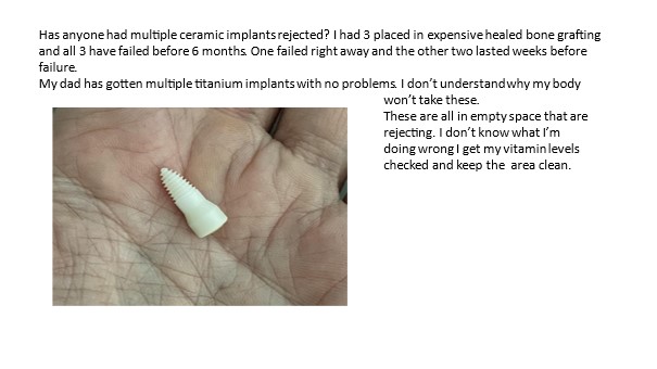
Patient 2: On Facebook a young patient reports:

Has anyone had multiple ceramic implants rejected? I had 3 placed in expensive healed bone grafting and all 3 have failed before 6 months. One failed right away and the other two lasted weeks before failure.

Question:

Is the grafting ready to hold the implants?

How to determine the bone density of grafting?

An ultrasound sonography with CaviTAU® might be the answer!

Call for ICOSIM study

The number of providers of bone replacement materials for the augmentation of reduced jaw conditions of various origins is increasing. This leads to increased statements by dental users and manufacturers about the success of the respective product, which is usually supported by local histological sections.

The success of an augmentation is consistently measured by the degree of ossification of the artificial augment. The success of implantation into the augmented replacement tissue depends on its osteoneogenic quality.However, it seems difficult to reliably determine the actual density ratios for dentists in practice with the X-ray methods available so far.

There is therefore both a diagnostic gap – simple measurement of bone density in the augment; and a therapeutic gap – the dentist cannot reliably determine a reliable time for implantation.

The aim of the study is therefore to measure the density of the augment using the medically approved and scientifically validated CaviTAU® ultrasound sonography procedure and thus obtain reliable statements about the degree of healing and implant readiness of the bone substitute. At the same time, a sample is taken and cytokine analysis of the augment takes place at the end stage of healing during implantation.


Sponsors of the study can be:

a) Dentists who want to check the clinical valence of their practice method for augmentation and want to present this to patients as a reference to their implantation expertise with scientific data material and publication.

b) Manufacturers of replacement materials who want to follow and present a retrospective course study of the healing degree of their product in cooperation with dentists they trust. They gain medically relevant statements about the quality of their product in comparison to the market offering.

If you are interested in participating, please contact me: drlechner@aol.com

2. Save the Date: 

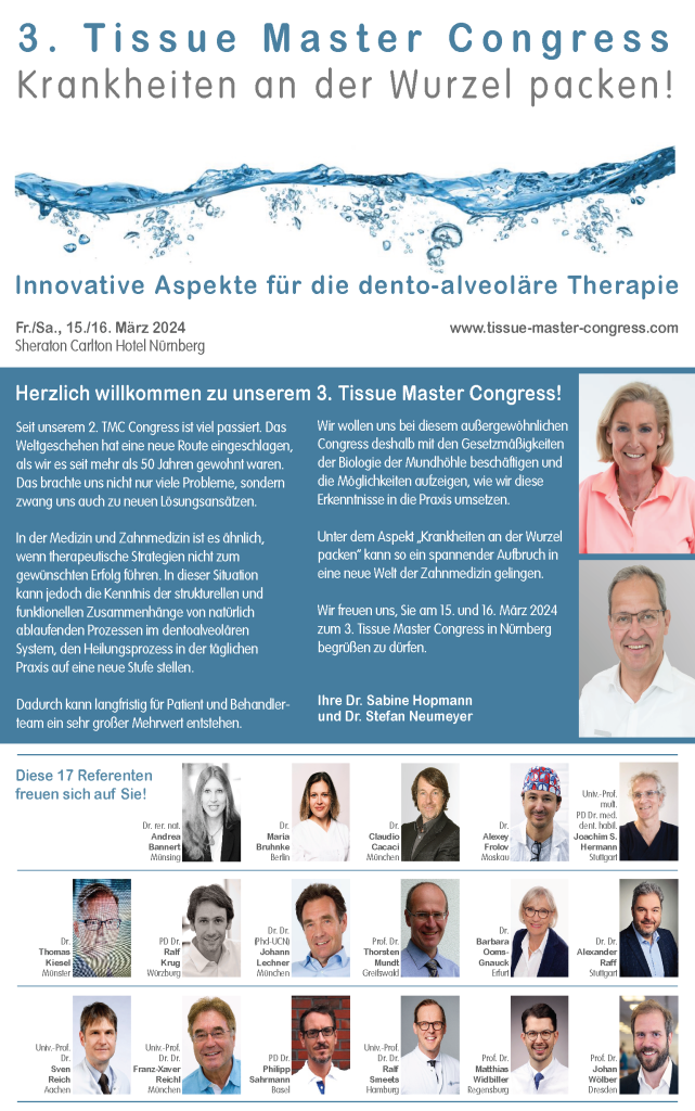
3. Tissue Master Congress “Innovative aspects for dento-alveolar therapy – tackling root diseases”

will take place from March 14-16, 2024 in Nürnberg

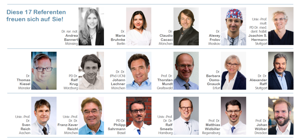
The event will feature a lecture by Hans Lechner on “Osteoimmunology and implant success – long-term immune sustainability of dental implants.” 

You can download the flyer or visit the congress website for more information and registration details: 


I look forward to seeing you in Nuremberg and remain with collegial greetings until the next ICOSIM Weekly.

Your Hans Lechner.

26. ICOSIM Weekly

1. This is how to handle radiation exposure responsibly
2. Save The Date: International Congress for Integrative Dentistry December 1st and 2nd, ’23

Dear friends of ICOSIM and
jawbone osteoimmunology,


It is recommended to read the current S2k guideline (long version) on radiation exposure DVT-justifying indication from both the perspective of patient protection and forensic aspects.

Here are four points from the perspective of radiation-free ultrasound sonography in dental, oral, and maxillofacial medicine for science-oriented ICOSIM members:


1.I. Dental digital volume tomography – DVT

AWMF registration number: 083-005 Status: December 2022 Valid until: December 2027

The implantological operator should be aware of the increased radiation exposure associated with DVT compared to two-dimensional imaging.

…However, three-dimensional imaging may still be justified for orofacial pathologies and diseases of the jawbone as well as a number of specific indications (European Commission 2012; Kapila and Nervina 2015; P. Jaju and S. Jaju 2015). The justifying indication requires the determination that the health benefit of the application outweighs the radiation risk. Other procedures with comparable health benefits that are associated with no or lower radiation exposure should be considered in the evaluation.

…Whereby the radiation exposure caused by an X-ray examination is to be limited as far as possible in accordance with the requirements of medical science (ALARA principle). When selecting the respective procedure, it should be taken into account that DVT application is associated with at least 10 times the effective dose compared to intraoral tube or bite-wing radiography.

Since there is still insufficient evidence for the use of DVT technology in childhood and adolescence for many questions, the indication must be carefully weighed on a case-by-case basis (European Commission 2004; European Commission 2012). A replacement production of a DVT recording exclusively for subsequent calculation of two-dimensional recordings (e.g. panoramic reconstruction or remote X-ray lateral recording) is not justified for radiation protection reasons.

In the application, the internationally established ALARA principle should generally be followed. Increasingly, the ALADA (as low as diagnostically acceptable) principle is also applied, which focuses on optimizing radiation dose in medical imaging, as proposed by the National Council on Radiation Protection and Measurements (NCRP) (White et al., 2012; Yeung, Jacobs, & Bornstein, 2019).

Risk assessments are difficult at low dose levels and are mainly based on the evaluation of cohorts of survivors of the atomic bombings in Japan in 1945 in combination with the internationally accepted “Linear No Threshold (LNT) assumption” in radiation protection (BEIR, 2006).

The estimated risk of dying from exposure to ionizing radiation from a DVT scan (risk of exposure-induced death (REID)(BEIR 2006)) is on average six cases per million (Pauwels et al., 2014), with women having a 40% higher risk than men on average (Pauwels et al., 2014). Another study calculated twice the risk for ten-year-olds compared to thirty-year-olds (Yeh & Chen, 2018).

In accordance with the optimization principle known in radiation protection, the possibilities for using dose-saving techniques, especially in the production of DVT images in children and adolescents as well as the S2k guideline “Dental digital volume tomography” long version as of December 2022© ARö, DGZMK 16 should be exhausted as far as possible (Federal Government of Germany 2018).

II. Bone density and DVT

In addition, it should be noted that structures located outside the image-generating FOV can lead to increased image noise due to the local tomography effect (Arai et al. 1999; Daatselaar, van der Stelt, and Weenen 2004; Katsumata et al. 2009).

For example, in peri-implantitis diagnosis, diagnostics of the immediate peri-implant environment (such as osseointegration of a dental implant) is only possible to a limited extent due to artifacts in DVT and CT (Draenert et al. 2007; Schulze, Berndt, and d’Hoedt 2010). To what extent DVT is possible against the background of existing imaging errors in the immediate vicinity of implant imaging cannot be reliably clarified based on current scientific data.


III. Is Hounsfield validated in DVT?

Currently, there are no randomized or controlled studies in patients that demonstrate the benefits of three-dimensional diagnostics with regard to the quality of surgical outcomes and/or the frequency of complications in implantology.

However, it should be noted that comparisons of noise between DVT devices or with CT are currently not valid, as the required calibration of Hounsfield units is not currently available on any DVT device (Blendl et al. 2012; Pauwels, Jacobs et al. 2015).

The gray values displayed in DVT are not standardized, unlike the values normalized over the Hounsfield scale in CT. Therefore, a quantitative use of gray values in DVT images, such as for bone density estimation, is not possible between different devices (Bornstein, W. Scarfe et al. 2014).


IV. Conclusion

a) Radiation exposure is harmful; its immunological safety is not proven. The Linear No Threshold (LNT) assumption applies internationally. There is no reliable “threshold” because the summation effect of radiation doses contradicts their health sustainability.

b) Image noise and imaging errors in the immediate implant environment limit DVT statements.

c) Hounsfield units in DVT are neither validated nor reliable.

d) Everyone should think further about their medical and forensic responsibility!


Therefore, the following principle applies, which you will also learn in training courses such as a CaviTAU® training: First OPG, then ultrasound examination, and only if the ultrasound examination is positive, then DVT due to radiation protection law (ALARA principle).
 

2. Save The Date 

Further education amidst a picturesque setting?
Plenty of sunshine instead of gray days?

Hans Lechner and other actors, such as the manufacturer CaviTAU®, are represented at the International Congress for Integrative Dentistry in Porto, Portugal with a workshop and lecture: ‘The smouldering fire in the jawbone – Systemic diseases and neglected RANTES inflammation detected by ultrasonography.’


To register and for more information, simply click on the button
to go to the congress website.


Until the next ICOSIM Weekly, I remain with collegial greetings.

Your Hans Lechner.

25. ICOSIM Weekly

1. Failed augmentation – A letter from a patient documents the need to use a device to measure bone density in augmentations

Dear friends of ICOSIM and
jawbone osteoimmunology,


 In this weekly, I present a case study of a patient that documents the need to use a device for measuring bone density in augmentations:

 
I had an implant from SDS, with internal sinus lift, inserted by a specialist about 2 years ago. After a healing time of 7.5 months, the implant was removed again, because it started to wobble 2 weeks after the crown was placed. The dentist pulled the implant out again.
 
After the implant removal, I suffered from dizziness for days. I also have joint pain since then, which I never had before. 
 
I am 58 years old and basically healthy, have good teeth (first denture) and have a
good immune system. My D3 level (87 ng/ml) and other nutrients are very well
present according to blood count.
Therefore, it is unclear why the implant did not hold.
 

My answer 1:


a) To your case:

The colleague works with his bone augmentations – like many other dentists – without knowing whether this artificial attempt was successful, because he has no device to determine the bone density of his augmentation so firmly that the inserted implants now hold or not.

With an ultrasound sonography, performable with a CaviTAU® device, he could have checked that and the implant would not have fallen out after loading (by the way, you are not an isolated case; the internet is full of such complaints).

Answer patient:In addition, I am still looking for the right way (implant and bone augmentation including material) and a specialist whom I can trust in this regard. My mother (81 years) got very many implants (no SDS) inserted within one year. She has been physically and mentally very bad since then. Meanwhile, there are expert opinions on her and she was advised to make claims for damages against this dentist.

My answer 2: 


b) The problem lies in the constant x-raying and trusting that x-rays reliably show the bone density in the jaw.

  1. BEFORE the implantation – then implants are inserted in not well healed jaws and the patient suffers from reactivated old inflammations (case of your mother).

  2. BEFORE the implantation in bone augmentation – then the dentist does not know whether this augmentation has already healed so firmly that the implant holds (your case).


c) To determine the bone or augmentation density in case a) and b), there is the possibility to use the – radiation-free (there is also a radiation protection law for dentists) – ultrasound sonography with the help of CaviTAU®  to avoid cases a) and b) in the interest of patient protection. 


Unfortunately, the above-mentioned patient is not an isolated case, as the internet in the USA shows:

Please think of the valuable application of a CaviTAU®-device for augmentations in volume IV, chapter 4.5: TAU for bone augmentation.

You can download a new flyer with information and ordering options here: :

New flyer for information and order here for DOWNLOAD


You can also find the volumes directly in our ICOSIM SHOP:

To the SHOP literature overview


I look forward to your numerous feedbacks on this completely unresolved problem in the entire implantation surgery.


With best ICOSIM regards

Your Hans Lechner

24. ICOSIM Weekly

1. New PubMed publication from CaviTAU® users
„ Reduction of Inflammatory RANTES/CCL5 Serum Levels by Surgery in Patients with Bone Marrow Defects of the Jawbone“
2. Training: Global cancer increase and what we can do about it

Dear friends of ICOSIM and jawbone osteoimmunology,

 
  1. I am pleased to share a new PubMed publication by CaviTAU® users and RANTES/CCL5 Jawbone Detox®remediators Reduction of Inflammatory RANTES/CCL5 Serum Levels by Surgery in Patients with Bone Marrow Defects of the Jawbone“ 

I am glad if you like the paper.

  1. On the not so unproblematic topic of CANCER, today I am putting together three scientific facts and – fourth – the conclusion from them on osteoimmunology of the jaw and CaviTAU®. That our approach to RANTES/CCL5 remediation and the detection of the sources in the jaw is correct, as shown by the above scientific publication.
  1. Startling scientific statistic: cancer cases in under-50s worldwide up nearly 80% in three decades, study finds


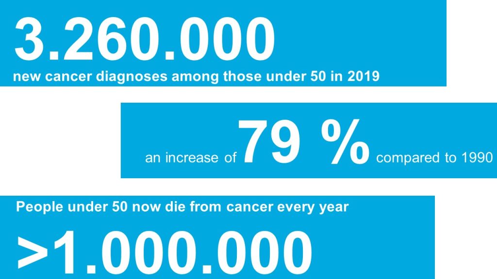
More than one million people under the age of 50 die from cancer each year, and the number is expected to increase by another 21% by 2030. The number of global cases of early-stage cancer has increased from 1.82 million in 1990 to 3.26 million in 2019, while cancer deaths among adults aged 40, 30 years or younger increased by 27%.

More than one million people under age 50 now die of cancer each year, the study found.

The authors of the study, published in BMJ Oncology, note that poor diet, alcohol and tobacco use, physical inactivity and obesity are likely contributing factors.

“Since 1990, incidence and deaths from early-stage cancers have increased substantially worldwide,” the report states. “Promoting healthy lifestyles, including a healthy diet, limiting tobacco and alcohol use, and adequate outdoor exercise, could reduce the burden of early-stage cancer.”

The recent study, led by the University of Edinburgh in Scotland and Zhejiang University School of Medicine in Hangzhou, China, was the first of its kind to examine the issue on a global scale and risk factors for younger adults. Most of the previous studies focused on regional and national differences. In this global study, researchers analyzed data from 204 countries on 29 cancer types.

In 2019, a total of 3.26 million new cancer diagnoses were made among those younger than 50 years, a 79% increase from 1990. Breast cancer was the most common cancer and the most common cause of death, with 13.7 and 3.5 cases per 100,000 of the global population, respectively.

In 2019, a total of 1.06 million people under the age of 50 died from cancer, a 27% increase from 1990. After breast cancer, most deaths were associated with cancers of the trachea, lung, stomach, and intestine.

Based on trends observed over the past three decades, researchers estimate that the number of new cancer cases and associated deaths worldwide will increase by another 31% and 21%, respectively, by 2030, with people in their 40s most at risk.

Dr. Claire Knight, a senior health information manager at Cancer Research UK who was not involved in the study, said it is not yet clear what is driving this trend and urged caution. “If people are concerned about their cancer risk, there are many ways to reduce it, such as not smoking, eating a balanced diet, getting plenty of exercise and staying safe in the sun.

  1. Fact #2: Cancer and RANTES / CCL5

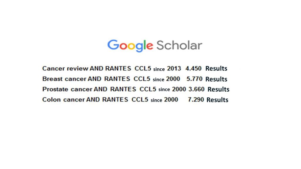
What does the scientific literature say about RANTES/CCL5 and tumors? Searching for “Cancer review … or Breast cancer …or Prostate cancer…or Colon cancer AND RANTES CCL5” in the science database GoogleScholar yields surprising results:
  1. Fact #3: RANTES / CCL5 and CaviTAU®

Research shows that conspicuous bone softening in the jaw (FDOJ) is a common, almost epidemic phenomenon in old extraction and implant sites. Please refer to:

  1. Lechner J, von Baehr V. RANTES and fibroblast growth factor 2 in jawbone cavitations: triggers for systemic disease? Int. Jour. of General Medicine; 2013:6 Pages 277 – 290, DOI: https://dx.doi.org/10.2147/IJGM.S43852
  2. Lechner J, von Baehr V. Hyperactivated Signaling Pathways of Chemokine RANTES/CCL5 in Osteopathies of Jawbone in Breast Cancer Patients—Case Report and Research. Breast Cancer: Basic and Clinical Research 2014:8 89–96 DOI. https://doi.org/10.4137%2FBCBCR.S15119
  3. Lechner J, Schmidt M, von Baehr V, Schick F. Undetected Jawbone Marrow Defects as Inflammatory and Degenerative Signaling Pathways: Chemokine RANTES/CCL5 as a Possible Link Between the Jawbone and Systemic Interactions?. J Inflamm Res. 2021;14:1603-1612. https://doi.org/10.2147/JIR.S307635
  4. Lechner J, Schulz T, Lejeune B, von Baehr V. Jawbone Cavitation Expressed RANTES/CCL5: Case Studies Linking Silent Inflammation in the Jawbone with Epistemology of Breast Cancer. Breast Cancer (Dove Med Press). 2021;13:225-240 https://doi.org/10.2147/BCTT.S29548

  1. The logic from #1 + #2 + #3 yields the ICOSIM proposal for the cancer question:

  1. Wouldn’t a systematic screening for “Silent Inflammation” in the jaw and thus for potentially cancer-causing RANTES/CCL5 “foci” radiation-free and non-invasive with CaviTAU® ultrasound sonography be a preventive and revolutionary addition to the smoke- and meat-free sedentary life?
  2. Wouldn’t the systematic search for the causes of cancer development with detection of “silent inflammation” in the jaw with radiation-free ultrasound be the more effective supplement to the much-discussed X-ray mammography?

With this I remain until the next ICOSIM-Weekly with collegial greetings.


Yours
Hans Lechner

23. ICOSIM Weekly

1. Immunological sustainability of titanium implants
2. Abstract for the article
3. Don´t miss the date:
Blood Concentrate Day Frankfurt 15. Sep. 2023

Dear friends of ICOSIM and jawbone osteoimmunology,


Has implantology already reached its scientific endpoint? In the daily routine of implanting is everything perfect or are we forgetting hidden stresses on the immune system?


1. A possible answer is given in this Weekly – By the abstract for the latest publication of our Munich Research Team MRT:

Immunological sustainability of titanium implants?
Osteoimmunology and osseointegration in dissolved titanium particles


Abstract: The bone-implant contact (BIC) is the essential prerequisite for successful osseointegration and implant stability. If this BIC does not occur on the entire surface of the implant, osseointegration is disturbed and local chronic inflammations in the sense of osteoimmunology can occur. These fatty-degenerative osteonecroses of the jawbone (FDOJ) endanger the immunological sustainability of the implantation; they harm the patient systemically by local RANTES/CCL5 overexpression at the implant-bone interfaces. Since no technique has yet been developed to visualize and verify whether bone or soft tissue is actually present around implants (Albrektsson 2023), these bone marrow defects at the BIC of implants have not been explored. A computerized transalveolar ultrasound system covers that diagnostic gap of standard X-ray techniques. A clinical case presentation demonstrates the immunological relevance of the problem and shows the solution using complementary dental sonography.


2. Curious? This is the PDF download of the original IMPLANTS article in English

Immunological sustainability of titanium implants? 
Osteoimmunology and osseointegration in dissolved titanium particles

3. Save the Date: International Blood Concentrate Day Frankfurt 15. Sep. 2023


Topic of the Dr. Lechner lecture:Bone Ultrasound Detection of Difficult to Diagnose Bone Affections after Extraction and/or Implantation”. 


Topic of the Dr. Lechner workshop:Between Osseointegration and Osteoimmunology – Practice of Ultrasound Sonography for Immunological Sustainability of Titanium and Ceramic Implants


I remain until the next ICOSIM Weekly with collegial greetings in preparation for the event in Bad Kissingen at the IGAF and the Blood Concentrate Day with Prof. Ghanaati in Frankfurt.


Yours,
Hans Lechner